
With the widespread adoption of low-dose CT screening, the detection rate of pulmonary nodules has increased significantly. Video-Assisted Thoracoscopic Surgery (VATS) has become the mainstream surgical approach for pulmonary nodule resection, offering advantages such as minimal invasiveness, enhanced safety, and faster postoperative recovery. However, for small-sized, deeply located nodules, intraoperative visualization and tactile localization are challenging. Without precise localization, the procedure may result in inaccurate resection margins, prolonged operative time, and in severe cases, conversion to open thoracotomy, increasing patient trauma and procedural risk.
To address the clinical challenges of intraoperative localization difficulty and prolonged lesion exploration, our R&D team has worked closely with surgeons to systematically evaluate the shortcomings of conventional localization products—such as a tendency to migrate or detach, and the inability to visualize nodule depth. Drawing on extensive clinical feedback, we conducted multiple rounds of optimization and rigorous validation, focusing on key performance parameters including anchoring force, flexibility, and safety. By overcoming numerous technical hurdles, we independently developed a four-prong anchor-based pulmonary nodule localization needle, providing a stable and reliable preoperative localization solution for precise VATS procedures.
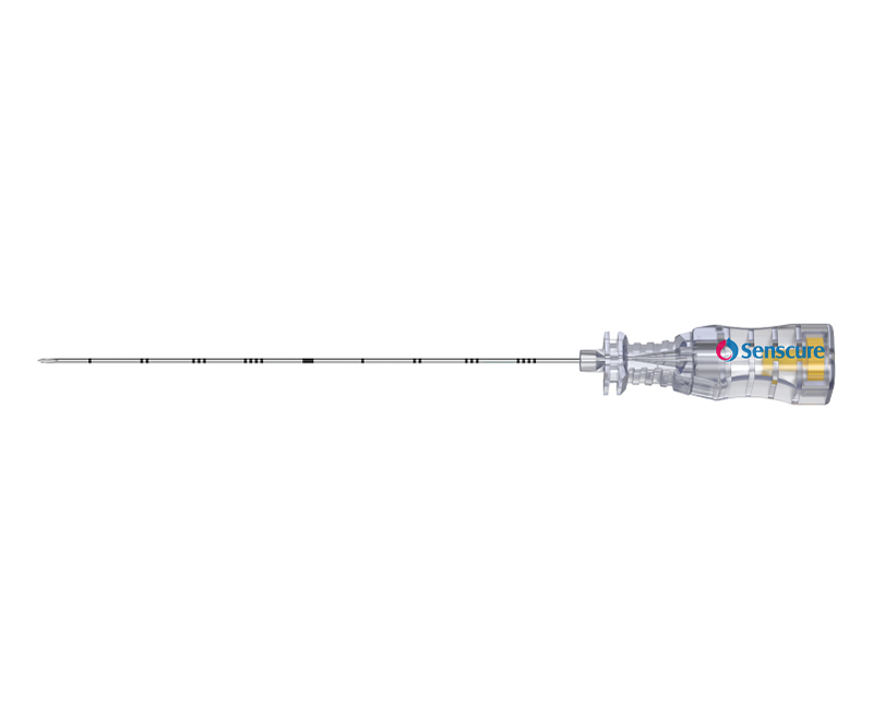
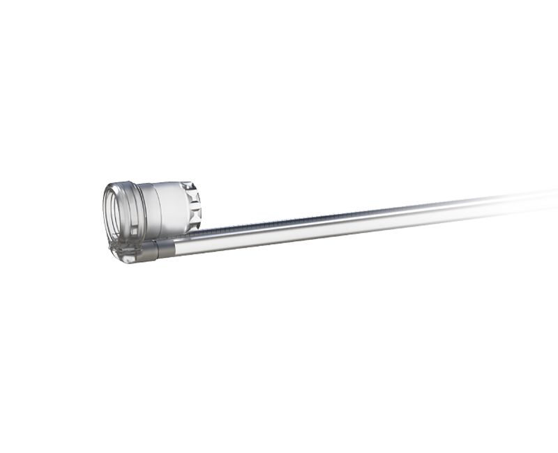
This product is a dedicated preoperative localization device for VATS pulmonary nodule surgery, designed for percutaneous placement under CT guidance. Its core structure consists of a nitinol memory-alloy four-prong anchor hook combined with a color-coded flexible localization thread. Preoperatively, the anchor hook, maintained in a retracted state, is precisely delivered to the periphery of the nodule. Upon deployment, it automatically expands to securely anchor the target tissue, leaving a visible localization marker. This fundamentally resolves the challenge of intraoperative localization for deeply located or small nodules, effectively avoiding blind resection and the risk of conversion to thoracotomy.
The product offers multiple core features and technical advantages. Its secure anchoring and controlled safety are ensured by the four-prong anchor hook, which provides stable fixation, while the blunt, rounded tip design effectively reduces the risk of complications such as lung injury, bleeding, and pneumothorax, enhancing overall safety. The flexible buffer and anti-migration design incorporates a flexible localization thread that absorbs intraoperative traction forces, significantly reducing the risk of anchor hook migration or dislodgement, ensuring reliable and stable positioning. The color-coded visual depth indication enables efficient localization, with segmented color markings that intuitively display nodule depth, eliminating visual blind spots during surgery and substantially shortening lesion exploration time, thereby improving procedural efficiency.
In clinical practice, accurate preoperative localization effectively resolves the positioning challenges of traditional VATS procedures, enabling precise targeting of lesions and avoiding excessive resection of healthy lung tissue, insufficient margins, or residual lesions. This maximizes preservation of lung function and improves long-term patient outcomes. Furthermore, the product offers a flexible time window for surgery scheduling, streamlines the surgical workflow, and significantly reduces the incidence of complications and conversion to thoracotomy. It comprehensively enhances the precision and safety of minimally invasive diagnosis and treatment for pulmonary nodules, representing a pivotal technology for early-stage pulmonary lesion management.

 |
| 
 +86-574-63729798
 overseas@senscure.net
 Block B, Building 3, No.777 Binhai 4th Road, Hangzhou Bay New District, Ningbo 315336, Zhejiang Province, China